|
|
|
 |
Summary
Anterior Cruciate Ligament Repair or ACL repairs or reconnects the torn ACL ligament. The ACL is one of the four ligaments that stabilize the knee. The ACL is the most common of the four knee ligaments to become damaged or torn.
The anterior cruciate ligament restrains forward motion of the shin bone. In other words, it prevents the shin from going past the knee. Without it the shin and knee are unstable. In actuality, the ACL needs to be replaced rather than repaired if it is torn.
Reasons to choose ACL repair:
- You have a complete or partial tear of the ACL
- You completed rehabilitation and your knee is still unstable
- You are active in sports or work and your knee is unstable
- Chronic ACL deficiency has become life altering
Length of the procedure: 1 to 2 hours
Hospital Stay:ACL repair is done arthroscopically and does not require a hospital stay.
Recovery before traveling home:Patients seeking and anterior cruciate ligament repair through medical tourism can return home within 24 to 48 hours in most cases.
Procedure Details
When your procedure begins you will be given general anesthesia or a spinal block. The surgeon will make 2 to 3 small incisions around the knee. Saline solution is pumped in one of the incisions to expand the area and make the structures of the knee more visible. A tiny camera is inserted in another so an image of the underlying structures can be transmitted for the surgeons viewing.
The surgeon will drill small holes in the surrounding bones of the upper and lower leg. These holes will be used to anchor the ligament graft that will repair/replace the damaged ACL. The graft ligament is pulled through two of the anchor holes and secured with screws or staples. The incisions are closed with sutures or tape.
After the Procedure
When the surgery is complete you will be moved to a recovery area to be monitored. Once you have recovered from the anesthesia you were given you will be released from care. It is best to take part in some type of professional physical therapy to ensure proper healing and full restoration of mobility. It takes between 6 to 12 weeks to return to sports and normal activities following the procedure.
Result
Anterior cruciate ligament repair is chosen because it enables injured individuals to return to normal activity. It stabilizes the knee and prevents further injury from occurring. Studies show that close to 90% of individuals who have their ACL repair achieve favorable results.
Risks and Complications
ACL surgery is generally a safe procedure, but as with any medical procedure there are some risks. The following are the most common risks associated with ACL repair:
- Allergy to anesthesia
- Breathing problems from anesthesia
- Numbness in scar
- Infection
- Knee structure damage
- Nerve damage
- Arterial damage
- Blood clots
- Loosening of the graft tendon
- Limited mobility
- Bleeding
- Weakness in the knee
- Pain
- Chronic pain
- Swelling
- Chronic inflammation
- Repeat Injury
Any concerns you have before or after your ACL repair procedure regarding these risks and complications should be discussed with your surgeon.
Contact your physician if:
- Bleeding is soaking through your bandages and pressure applied to the area does not control the bleeding
- Pain does not subside with pain medication
- Swelling or pain in the calf develops
- Your toes or feet are cool to the touch and or darker than normal in color
- Redness of the incision develops
- Swelling of the incision develops
- The incision is throbbing or has localized pain
- The incision begins to ooze or has a yellowish discharge
- Your temperature is above 101 degrees and does not reduce when you take Tylenol or ibuprofen
|
| If you are considering argon-helium knife cryosurgery therapy in China and would like to get know more information about argon-helium knife cryosurgery therapy, please complete the inquiry form or email us hq@1uchina.com |
|
 
Definition
Achilles tendonitis causes inflammation and degeneration of the achilles tendon. The achilles tendon is the large tendon located in the back of the leg that inserts into the heel. The pain caused by achilles tendonitis can develop gradually without a history of trauma. The pain can be a shooting pain, burning pain, or even an extremely piercing pain. Achilles tendonitis should not be left untreated due to the danger that the tendon can become weak and ruptured.
Achilles Tendonitis is aggravated by activities that repeatedly stress the tendon, causing inflammation. In some cases even prolonged periods of standing can cause symptoms. It is a common problem often experienced by athletes, particularly distance runners. Achilles Tendonitis is a difficult injury to treat in athletes due to their high level of activity and reluctance to stop or slow down their training.
Individuals who suffer from achilles tendonitis often complain that their first steps out of bed in the morning are extremely painful. Another common complaint is pain after steps are taken after long periods of sitting. This pain often lessens with activity.
Cause
There are several factors that can cause achilles tendonitis. The most common cause is over-pronation. Over-pronation occurs in the walking process, when the arch collapses upon weight bearing, adding stress on the achilles tendon.
Other factors that lead to achilles tendonitis are improper shoe selection, inadequate stretching prior to engaging in athletics, a short achilles tendon, direct trauma (injury) to the tendon, and heel bone deformity.
Treatment and Prevention
Athletes, particularly runners, should incorporate a thorough stretching program to properly warm-up the muscles. They should decrease the distance of their walk or run, apply ice after the activity and avoid any uphill climbs. Athletes should use an orthotic device, heel cup, or heel cradle for extra support.
A heel cup or heel cradle elevates the heel to reduce stress and pressure on the achilles tendon. The device should be made with light-weight, shock absorbing materials. An orthotic device can be used to control over-pronation, support the longitudinal arch, and reduce stress on the achilles tendon.
If the problem persists, consult your foot doctor.
|
| If you are considering argon-helium knife cryosurgery therapy in China and would like to get know more information about argon-helium knife cryosurgery therapy, please complete the inquiry form or email us hq@1uchina.com |
|
What is ankle joint replacement?
Ankle joint replacement (also called ankle arthroplasty) is a surgical procedure to replace damaged parts of the ankle joint using artificial replacements (prostheses).
Who can benefit from ankle joint replacement surgery?
Most patients who undergo ankle joint replacement surgery suffer from arthritis of the ankle, most commonly osteoarthritis and rheumatoid arthritis. Another common reason may include injury to the ankle. Individuals may be aware of severe pain, stiffness and mobility problems and ankle joint replacement surgery may be the best solution to remedy this.
Procedure
Standard ankle joint replacement procedure is usually carried out under a general or spinal anesthetic. An incision is made on the front of the ankle, and any overlying tendons, blood vessels and nerves are moved out of the way. The ankle joint is then opened through a second incision, before the surgeon prepares the ankle joint by cutting and shaping the bone to fit the new prostheses. The prostheses consist of two parts: the ‘tibial’ component that replaces the socket (tibia), and the ‘talar’ component that replaces the upper part of the bone in the foot (talus). These are made of a mixture of plastic and metal. Once the replacement bone and socket are fitted, they are tested for friction and mobility. To help prevent any loosening of the prostheses, bone is grafted between the tibia and the bone that sits next to it – the fibula. Finally, the prostheses are then screwed into place. After the surgeon has finished implanting the prostheses, the incisions are closed using sutures or staples. A splint is then bandaged onto the ankle to help keep the new joint in place during the healing period.
Recovery
Recovery following ankle joint replacement surgery will take some time. You will need to keep your foot elevated and need to avoid putting any weight on the ankle for about six weeks. A physiotherapist will help you to strengthen the ankle and regain mobility through specific exercises, as well as show you how to use a walker or crutches. You can take painkillers or will be prescribed medication to help alleviate any pain. Returning to work will depend on the nature of your job, and your physiotherapist and surgeon will talk you through this, as well as when you can resume normal activities (including driving).
Risks
Risks surrounding ankle joint replacement surgery include infection, bleeding, loosening of the prostheses, damage to blood vessels and nerves, skin loss surrounding the ankle, dislocation, persistent pain and a noticeable different in leg length.
|
| If you are considering argon-helium knife cryosurgery therapy in China and would like to get know more information about argon-helium knife cryosurgery therapy, please complete the inquiry form or email us hq@1uchina.com |
|
Arthroplasty is the operation for construction of a new movable joint. It is not applicable to every joint: in practice, its use is almost confined to the shoulder, the elbow, the hip, the knee, certain joints in the hand, and the metatarso-phalangeal joints in the foot.
Indications
The indications for arthroplasty are not well defined, for there is considerable diversity of opinion among different surgeons. Broadly, it has a use in the following conditions:
- advanced osteoarthritis or rheumatoid arthritis with disabling pain, especially in the shoulder, elbow, hip, hand and metatarso-phalangeal joints;
- for the correction of certain types of deformity (especially hallux valgus);
- quiescent tuberculous arthritis especially of the elbow or hip;
- certain ununited fractures of the neck of the femur. It will be released that in several of these conditions arthroplasty is an alternative to arthrodesis.
Methods of arthroplasty
Three methods are in general use:
- excision arthroplasty;
- half-joint replacement arthroplasty;
- total replacement arthroplasty. Each has its merits; disadvantages and special applications.
Excision arthroplasty. In this method one or both of the articular ends of the bones are simply excised, so that a gap is created between them. The gap fills with fibrous tissue, or a pad of muscle or other soft tissue may be sewn in between the bones. By virtue of its flexibility the interposed tissue allows a reasonable range of movement, but the joint often lacks stability. The method is applicable to all the joints for which arthroplasty is practicable except the knee and ankle. It is used most commonly at the metatarso-phalangeal joint of the great toe, in the treatment of hallux valgus and hallux rigidus. At the hip it may be used as a salvage operation after failed replacement arthroplasty.
Three methods of arthroplasty, as exemplified at the hip. Excision arthroplasty. Note the interposed soft tissue. Half-joint replacement arthroplasty: the femoral head is replaced by a metal prosthesis. Figure I5 Total replacement arthroplasty. The femoral head is replaced by a metal prosthesis and the acetabulum by a plastic socket. Both are held in place by acrylic filling compound or 'cement'.
Half-joint replacement arthroplasty
In half-joint replacement arthroplasty one only of the articulating surfaces is removed and replaced by a prosthesis of similar shape. The prosthesis is usually made from metal (as in replacement of the femoral head), occasionally from silicone rubber (as in replacement of A carpal bone); and when appropriate it may be fixed into the recipient bone with acrylic filling compound or cement'. The opposing, normal articulating surface is left undisturbed. The technique has its main application at the hip, where prosthetic replacement of the head and neck of the femur is commonly practised for femoral neck fracture in the elderly. It has rather a limited use elsewhere, an example being the replacement of the lunate bone by a silicone-rubber prosthesis in Kienbock's disease.
Total replacement arthroplasty. In this technique both of the opposed articulating surfaces are excised and replaced by prosthetic components. In the larger joints one of the components is normally of metal and the other of high-density polyethylene, and it is usual for both components to be held in place by acrylic 'cement'. In small joints such as the metacarpo-phalangeal joints a flexible one-piece prosthesis made from silicone rubber may be used.
Total replacement arthroplasty has proved very successful at the hip and to a lesser extent at the knee. It has been extended, so far with only moderate success, to many other joints including the shoulder, elbow, ankle, metacarpo-phalangeal joints and metatarso-phalangeal joints. A disadvantage which applies also to half-joint replacement arthroplasty-is that there is a tendency for the prosthesis to work loose after a variable time that cannot be predicted. A well-fitted replacement joint may, however, give good service for many years, especially in the case of the hip.
|
| If you are considering argon-helium knife cryosurgery therapy in China and would like to get know more information about argon-helium knife cryosurgery therapy, please complete the inquiry form or email us hq@1uchina.com |
|
Arthroscopic Subacromial Decompression(ASD) |
Arthroscopic subacromial decompression may be a difficult technique to learn; good equipment therefore is essential. Everything must be done under direct vision, and hence control of bleeding is an essential requirement. This may be achieved in one of two ways.
- Increase of intrabursal fluid pressure: this increases postoperative swelling, however.
- Coagulation diathermy: Initially, use of diathermy was difficult because the inflow fluid had to be changed from saline to either water or glycine. However, commercially available point source diathermy probes are available, and these do not require change of fluid and can be used in the presence of saline.
Procedure
1. Arthroscopic glenohumeral exam. Standard posterior portal 1.5-2.0 cm inferior, 1 cm medial to the posterolateral corner of the acromion directing cannula toward the coracoid tip anteriorly. High anterior portal at anterolateral corner of acromion inside- out with trocar or outside-in with 18 gauge needle.
2. Remove scope and redirect through the same posterior skin incision just at inferior edge of the posterior acromion. Direct cannula anteriorly along the roof of the acromion (inferior surface).
3. Enter bursal chamber under anterior one half of the acromion. Direct blunt trocar /cannula out anteriorly through the previous high anterior portal and retrograde an outflow cannula back into the subacromial space at the anterolateral corner of the acromion.
4. Incise a lateral working portal 2.5-3.0 cm from lateral border of the acromion slightly anterior to the midpoint. Angle slightly upward so shaver/ablater does not impinge on the lateral cortex.
5. Define the anterior one half of the bony acromion with shaver and cautery/ablation from the lateral portal. Debride enough bursa to visualize well. May need to remove some of the “posterior bursal curtain”.
6. Ablate or transect coracoacromial ligament using the cautery unit or shaver. Have cautery ready for the acromial branch of the thoraco-acromial artery.
7. Resect the anterior 3-4mm of acromial bone from anterolateral corner toward the A-C joint using the burr in the lateral portal. Don’t enter the A-C joint if not planning a Mumford. (Figure 1)
8. Thin down lateral border of the anterior ½ acromion starting at the anterolateral corner and proceeding posteriorly tapering to the midpoint. (Figure 1)
")
Figure 1. Remove anterior 3-4 mm of acromion from the anterolateral corner to the AC joint, and thin down the lateral border of the anterior ½ of the acromion, tapering posteriorly.9. Place scope in lateral portal. View arch of acromion.
10. Introduce burr through posterior portal directly on undersurface of posterior ½ of the acromion.
11. Advance burr anteriorly on posterior “cutting block”. Use a sweeping motion medially to laterally as move anterior. (Figure 2)
")
Figure 2. The burr is advanced anteriorly using the posterior half of the acromion as a "cutting block" to remove the bone with a sweeping motion medially to laterally.12. Resect remaining anterior hook of the acromion. (Figure 3)Take care not to advance burr anteriorly into deltoid fibers or fascia.
")
Figure 3. The anterior hook of the acromion is resected and the undersurface flattened in the sagittal plane.13. Flatten the underside of the acromion in the sagittal plane. (Figure 3) Must know the sagittal acromial morphology and thickness preoperatively to avoid excessive resection. (Figure 4)
12. Resect remaining anterior hook of the acromion. (Figure 3)Take care not to advance burr anteriorly into deltoid fibers or fascia.
")
Figure 4. Determine shape and thickness of acromion preoperatively to determine if “cutting block” technique or minimal anterior hook resection is warranted.")
14. Place scope posteriorly.
15. View the acromion. Insure it is flat in the medial-lateral plane. If have any residual bone along lateral acromion, use burr from the lateral portal to complete lateral acromial resection. (Figure 5)
")
Figure 5. The scope is placed posteriorly to view the acromion and insure that it is flat in the mediolateral plane.16. Verify appropriate resection – from at least 2 separate portals: enlarge portal and palpate with gloved finger if unsure.
17. Reduce flow pressure. Use the electrocautery unit to obtain hemostasis.
|
| If you are considering argon-helium knife cryosurgery therapy in China and would like to get know more information about argon-helium knife cryosurgery therapy, please complete the inquiry form or email us hq@1uchina.com |
|
Articular Cartilage Surgery Knee Or Ankle |
What is Articular Cartilage Restoration?
Articular cartilage is the smooth, white tissue that covers the ends of bones where they come together to form joints. Healthy cartilage in our joints makes it easier to move. It allows the bones to glide over each other with very little friction.
Articular cartilage can be damaged by injury or normal wear and tear. Because cartilage does not heal itself well, doctors have developed surgical techniques to stimulate the growth of new cartilage. Restoring articular cartilage can relieve pain and allow better function. Most importantly, it can delay or prevent the onset of arthritis.
Surgical techniques to repair damaged cartilage are still evolving. It is hoped that as more is learned about cartilage and the healing response, surgeons will be better able to restore an injured joint.
Preparation of Surgery
What should I expect before surgery?
Before surgery, your surgeon will need to find out as much as possible about your knee. In addition to your physical exam, you will need more X-rays and possibly other imaging tests, such as magnetic resonance imaging (MRI) and bone scans. Your surgeon may also need to use an arthroscope (discussed later) to check the lesion's location, size, and depth.
Surgical Procedure
What happens during surgery?
Many types of surgery have been developed for fixing articular cartilage injuries in the knee. When the decision is made to go ahead with surgery, the surgeon will consider whether to do a procedure to restore or to repair the cartilage. A reparative surgery can help fill in the lesion, but it doesn't completely restore the actual makeup and function of the original cartilage. (Sometimes that simply isn't possible given the amount of damage in the knee.) Reparative procedures may provide pain relief and improve knee motion and function.
Your surgeon would ideally like to help your knee return to its natural state, with full function and no pain. This requires restorative surgery, meaning that the end result is a lesion filled to the full depth by tissue identical to the original. Surgeons rely on some fairly new procedures to substitute or replace the original cartilage. One method is to transplant cartilage and underlying bone from a nearby area in the knee joint. Another method is to take some chondrocytes (the primary cells of cartilage) from your knee cartilage, grow them in a laboratory, and then use the newly grown tissue to fill in the lesion at a later date.
The final decision about which surgery to use will be based on your specific injury, age, activity level, and the overall condition of your knee.
Types of Surgery
In general, recovery from an arthroscopic procedure is quicker and less painful than a traditional, open surgery. Your doctor will discuss the options with you to determine what kind of procedure is right for you.
The most common procedures for cartilage restoration are:
- Microfracture
- Drilling
- Abrasion Arthroplasty
- Autologous Chondrocyte Implantation
- Osteochondral Autograft Transplantation
- Osteochondral Allograft Transplantation
After Surgery
What happens after surgery?
After surgery, patients go to the post-anesthesia care unit (PACU) for specialized care until they awaken. Then they are either transferred to the nursing unit or released from the hospital. Many of the procedures for treating articular cartilage are done on an outpatient basis, meaning you can leave the hospital the same day.
Since surgeons use different methods when treating articular cartilage lesions in the knee, the instructions patients need to follow after surgery depend on the surgeon and the way the surgery was done.
Complications
What might go wrong?
As with all major surgical procedures, complications can occur. This document doesn't provide a complete list of the possible complications, but it does highlight some of the most common problems. Some of the most common complications following articular cartilage surgery are
- anesthesia complications
- thrombophlebitis
- infection
- hardware failure
- failure of surgery
|
| If you are considering argon-helium knife cryosurgery therapy in China and would like to get know more information about argon-helium knife cryosurgery therapy, please complete the inquiry form or email us hq@1uchina.com |
|
Artificial Disc Replacement (ADR) |
Overview
Artificial Disc Replacement (ADR), or Total Disc Replacement (TDR), is a type of arthroplasty. It is a surgical procedure in which degenerated intervertebral discs in the spinal column are replaced with artificial devices in the lumbar (lower) or cervical (upper) spine. The procedure is used to treat chronic, severe low back pain and cervical pain resulting from degenerative disc disease.
Artificial disc replacement has been developed as an alternative to spinal fusion, with the goal of pain reduction or elimination, while still allowing motion throughout the spine. Another possible benefit is the prevention of premature breakdown in adjacent levels of the spine, a potential risk in fusion surgeries.
How a Disc Is Replaced
The technique to install an artificial disc (whether in the neck or low back) is routine and safe. In both traditional disc surgery and artificial disc replacement the procedure begins by removing the gelatinous disc between the vertebrae.
- Once the disc is removed, two metal plates are pressed into the bony endplates above and below the space now vacated by the disc.
- Metal spikes hold these plates in place on the bone.
- Eventually bone will grow over and around the metal plates.
- The patient's own body weight compresses the spacer after the surgery is complete.
A plastic spacer made of a polyethylene core is put between the plates. The device allows for six degrees of freedom.
Recovery
Recovery from artificial disc replacement and care afterwards are much like that for other anterior approaches to lumbar spine surgery. In some cases, recovery is faster than for a traditional fusion surgery. There is less pain from the procedure and fewer complications in general. The materials used in artificial disc replacements are similar the materials used in routine hip and knee replacement surgery. The materials are designed not to cause sensitivities once in the body.
|
| If you are considering argon-helium knife cryosurgery therapy in China and would like to get know more information about argon-helium knife cryosurgery therapy, please complete the inquiry form or email us hq@1uchina.com |
|
Summary
A bunionectomy is a procedure that excises or removes bunions. Bunions form between the joint at the base of the big toe. They occur as a result of inflammation that is typically caused by shoes that are too tight or fit poorly, combined with the joint being overly mobile.
When these two factors combine a lump develops on the side of the joint. Overtime, this causes the big toe to push towards the second toe. This may also lead to new bone growth or a bone spur. The bunion is made up of soft tissue, and sometimes a bone spur.
You may consider a bunionectomy if:
- Conservative treatment has failed to correct the problem
- Your bunion has caused a foot deformity
- Intense pain has led to difficulty walking or performing other activities
- Your bunion has caused you to become unstable when walking
Length of the procedure: 1 hour
Hospital Stay:Depending on the method of surgery used and the severity of your problem you will be hospitalized for a maximum of 24 hours following the procedure, if at all.
Recovery before traveling home:Patients seeking a bunionectomy through medical tourism can return home within 24 to 48 hours in most cases.
Procedure Details
Your surgery will begin with the administration of anesthesia. Depending on what type of surgical method your doctor has chosen you will be given an ankle block, general anesthesia or a spinal block. Then, the area will be prepped with an antiseptic liquid.
During the surgery an incision will be made directly on the swollen area of your foot. The surgeon will remove the lump. They may also need to reposition the big toe. If this is necessary another incision may be required. If damage to the joint is present the surgeon may attach the bones of the foot with screws, wires or a metal plate. In some cases the joint may need to be replaced with an artificial joint.
After the Procedure
After the procedure your incisions will be closed with sutures or tape. The surgical wound will be wrapped in a compression dressing. The compression dressing will help maintain proper alignment of the bones and reduce swelling.
After the anesthesia has worn off in most cases you will be released from care. You will need to elevate your foot for at least 24 hours following the procedure. It will be at least 6 to 8 weeks before you can return to normal activity. Your surgeon may require you to wear a special orthopedic shoe or boot during this time. These orthopedic devices will help accommodate healing and provide you stability during the healing process.
Result
About 85% to 95% of patients consider their bunionectomy procedure successful. However, results depend largely upon a number of factors including age, severity of deformity, and the patients adherence to their surgical recovery instructions. Many patients also use orthotics after they heal from the procedure to keep their feet properly aligned and to prevent another bunion from forming.
Risks and Complications
As is with any surgery there are certain risks associated with a bunionectomy procedure. However, it is important to note that complications arise in less than 10% of bunionectomy patients. The following is a list of the most common risks and complications:
- Infection
- Pain
- Nerve damage
- Reoccurring bunion
- Reaction to anesthesia
Contact your physician if:
- Fever of 100.5 or above occurs and is uncontrolled by Tylenol or ibuprofen
- You have chills
- Pain is constant or increases
- There is redness of warmth in the surgical area
- Swelling develops in the calf
- Water or moisture causes the surgical dressing to come off
- The surgical dressing becomes bloody
|
| If you are considering argon-helium knife cryosurgery therapy in China and would like to get know more information about argon-helium knife cryosurgery therapy, please complete the inquiry form or email us hq@1uchina.com |
|
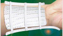
Summary
Carpal tunnel release is a surgery that is designed to treat individuals with carpal tunnel syndrome. This surgery is done on an outpatient basis. During the surgery patients are awake, but given anesthesia to block pain and encourage relaxation.
Carpal tunnel syndrome is caused by narrowing of the carpal tunnel passage. The narrowing of the passage leads to compression or pinching of the nerve. This causes pain and weakness in the hand and wrist.
Carpal tunnel release is usually performed on patients who have achieved little to no relief after being treated with non-surgical methods. A test called an EMG is done to diagnose carpal tunnel syndrome. Once diagnosis is confirmed, surgery should be scheduled promptly to prevent nerve damage.
Length of the procedure: 1 hour or less.
Hospital Stay:This is an outpatient procedure.
Recovery before traveling home:Patients seeking carpal tunnel release through medical tourism can return home within 24 in most cases.
Procedure Details
Your surgery begins with the administration of anesthesia to block pain and promote relaxation. The area will be prepped with a sterile antiseptic liquid. Then, an incision usually about an inch in length is made on the palm of the hand.
After the initial incision is made the surgeon will carefully expose the carpal tunnel by making an incision through the connective tissue immediately below the skin’s surface. When they have the carpal tunnel in sight they will release it by cutting it with a scalpel or scissors.
To finish the procedure the surgeon will stitch the skin together. The carpal tunnel is left divided. However, the two ends of the carpal tunnel will fill in with scar tissue over time.
After the Procedure
After the procedure your wrist will be wrapped in bandages and a splint. The splint and bandages will need to remain in place for about a week’s time. You may have some discomfort, but you will be given a prescription for pain medication to help relieve any discomfort.
One week after your carpal tunnel release surgery you will need to start performing motion exercises to promote healing and ensure returned function. Your doctor will recommend certain motion exercises or they may prescribe professional physical therapy. Depending on the extent of your carpal tunnel syndrome it can take a few weeks to an entire year to completely recover from the procedure.
Result
The prognosis for carpal tunnel release is very good. This procedure can effectively get rid of the pain, tingling, numbness and muscle weakness caused by carpal tunnel syndrome. In fact, nearly 85% of patients who have this surgery experience relief from their symptoms. Many also experience increased strength and restored function.
Risks and Complications
As is with any surgery there are risks associated with carpal tunnel release. However, this is considered a low risk and safe procedure. The following is a list of the most common risks and complications:
- Infection
- Pain
- Incision pain
- Scar sensitivity
- Arterial damage
- Inflammation
- Stiffness
- Hand or wrist weakness
- Injury to the median nerve
- Nerve damage
- Need for repeat repair caused by improper healing
- Scar tissue formation
Any concerns you have regarding these risks before or after the procedure should be discussed with your surgeon promptly.
Contact your physician if:
- Swelling increases
- Pain increases
- Pain is not relieved by medication
- Bleeding is excessive or uncontrolled
- Bright red bleeding comes from incision
- The incision feels warm or has a warm sensation
- The incision or surrounding are becomes red
- Discharge accompanied by an odor comes from the incision
- Nausea or vomiting develops
- Fever is uncontrolled with Tylenol or ibuprofen
- Dizziness develops
- Headache develops
- Muscle aches accompanied with chills
|
| If you are considering argon-helium knife cryosurgery therapy in China and would like to get know more information about argon-helium knife cryosurgery therapy, please complete the inquiry form or email us hq@1uchina.com |
|
Cervical Discectomy (endoscopic) |
For some people with chronic neck pain or other disorders that affect the cervical spine, an anterior cervical discectomy may be needed. The following information discusses this common surgical procedure.
The Cervical Spine
The human spine is divided into three sections:
- the cervical spine or neck (which is made up of 7 vertebrae)
- the thoracic spine (which is made up of 12 vertebrae)
- the lumbar spine or low back (which consists of 5 vertebrae)

The cervical spine begins at the base of the skull and supports the weight of the head. The spinal cord runs from the brain down through the cervical spine, controlling the function of the body's organs and limbs. In between each of the 7 vertebrae of the cervical spine are soft pads or discs which act as shock-absorbers and allow for bending and movement of the head. Each disc is made up of two parts, a soft center called the nucleus and a tough outer band called the annulus.
Cervical Pain
Millions of people suffer from pain in their necks or arms. A common cause of cervical pain is a rupture or herniation of one or more of the cervical discs. This happens when the annulus of the disc tears and the soft nucleus squeezes out. As a result, pressure is placed on the nerve root or the spinal cord and causes pain in the neck, shoulders, arms and sometimes the hands. Cervical disc herniations can occur as a result of aging, wear and tear, or sudden stress like from an accident.

Most cases of cervical pain do not require surgery and are treated using non-surgical methods such as medications, physical therapy and/or bracing. However, if patients experience significant pain and weakness that does not improve, surgery may be necessary.
Surgical Technique
An anterior cervical discectomy is the most common surgical procedure to treat damaged cervical discs. Its goal is to relieve pressure on the nerve roots or on the spinal cord by removing the ruptured disc. It is called anterior because the cervical spine is reached through a small incision in the front of the neck (anterior means front). During the surgery, the soft tissues of the neck are separated and the disc is removed. Sometimes the space between the vertebrae are left open. However, in order to maintain the normal height of the disc space, the surgeon may choose to fill the space with a bone graft.
A bone graft is a small piece of bone, either taken from the patient's body (usually from the pelvic area) or from a bone bank. This piece of bone fills the disc space and ideally will join or fuse the vertebrae together. This is called fusion. It usually takes a few months for the vertebrae to completely fuse. In some cases, some instrumentation (such as plates or screws) may also be used to add stability to the spine.
After Surgery
Patients will feel some pain after surgery, especially at the incision site. Pain medications are usually given to help control the pain. Upon a physician's direction, moist heat and frequent repositioning can also provide some relief. While tingling sensations or numbness is common, and should lessen over time, they should be reported to the doctor. Most patients are up and moving around within a few hours after surgery. In fact, this is encouraged in order to keep circulation normal and avoid blood clots.
However, most patients need to remain in the hospital, gradually increasing the amount of time they are up and walking, before they are discharged. Prior to discharge the doctor will provide the patient with careful directions about activities that can be pursued and activities to be avoided. Often patients are encouraged to maintain a daily low-impact exercise program. Walking, and slowly increasing the distance each day, is the best exercise after this type of surgery. Some discomfort is normal, but pain is a signal to slow down and rest.
Signs of infection like swelling, redness or draining at the incision site, and fever should be checked out by the surgeon immediately. Keep in mind, the amount of time it takes to return to normal activities is different for every patient. Discomfort should decrease a little each day. Increases in energy and activity are signs that recovery is going well. Maintaining a healthy attitude, a well-balanced diet, and getting plenty of rest are also great ways to speed up recovery.
|
| If you are considering argon-helium knife cryosurgery therapy in China and would like to get know more information about argon-helium knife cryosurgery therapy, please complete the inquiry form or email us hq@1uchina.com |
|
Summary
THR or total hip replacement is a common surgical procedure. The procedure rebuilds a deteriorated or damaged hip joint to make it more functional. During the procedure the damaged joint is replaced with an artificial prosthesis.
Signs that a hip replacement may benefit you:
- Hip pain is limiting your daily activities including walking and bending.
- Hip pain persists when you are resting during the day or night.
- Stiffness in the hip has reduced your ability to move or lift your leg.
- Anti-inflammatory drugs or glucosamine sulfate do not provide relief.
- Hip medications are causing harmful or unpleasant side effects.
- Treatments like physical therapy or using a cane do not relieve your pain.
Length of the procedure: 1 to 3 hours
Hospital Stay:4 to 10 days
Recovery before traveling home:When seeking a total hip replacement via medical tourism you can expect to travel in 7 to 10 days.
Procedure Details
When you arrive for your procedure you will be prepped for surgery and administered anesthesia. During the procedure your surgeon will remove diseased cartilage and bone first. They will also remove the ball and socket of the hip.
The ball and socket of the hip is then replaced with a metal ball and stem that is positioned inside the femur. Cement is used to attach this prosthesis to the femur. In some cases the prosthesis is placed without cement. This prosthesis has microscopic pores that connect to the femur bone over time.
After the Procedure
When the procedure is complete you will be in recovery for about 2 hours. You will be bandaged for about 24-48 hours. IV antibiotics will be administered to prevent infection, along with pain medication to ensure your comfort level. Blood thinners will be given to prevent clot formation.
Dislocation is common after total hip replacement. Certain measures will be taken to prevent this for the first 6 to 8 weeks after the procedure. These Measures include:
- placing one pillows between your legs
- avoiding crossing your legs
- not bending forward 90 degrees
- using a higher toilet seat
- not letting your knees and/or toes turn in
You will start physical therapy shortly after your procedure and continue with it for several weeks. Exercise will be prescribed for you to do independently and with a physical therapist. You will also use crutches or a walker for several weeks.
Your physical therapy regimen should include:
- Returning to sitting, standing, and walking up and down stairs
- Exercises several times a day that are designed to restore movement
- Exercises several times a day that are designed to strengthen your hip joint
- A graduated walking program that begins in your home and moves outside
- A walking program that slowly and steadily increases your mobility and endurance
- Returning to other normal household activities
Result
Most patients resume normal activities about three months after their procedure. Total hip replacement can help reduce pain, increase mobility and get you back to enjoying normal activities. The longevity of prosthetic hips varies, but can range from 10 to 25 years.
Risks and Complications
As with any surgery, there are certain risks and complications associated with total hip replacement including:
- Blood Clots
- Infection
- Dislocation
- Extra Bone Formation
- Femur Fracture
- Pain
- Stiffness
- Shortening of the Leg
- Arterial Injury
- Bleeding
- Allergy to Prothesis
- Allergy to Anesthesia
- Fat Embolism
It is important to discuss any concerns you have about the risks and complications with your surgeon before and after your total hip replacement.
Contact your physician if:
- You develop a persistent fever (higher than 100°F orally)
- Have excessive shaking chills
- You notice increasing redness, tenderness, or swelling of the hip wound
- Drainage from the hip wound occurs
- You have increasing hip pain with both activity and rest
- Pain in your calf and leg develops that is unrelated to your incision
- Tenderness or redness of your calf begins
- Your thigh, calf, ankle, or foot begin to swell
- Shortness of breath occurs
- Chest pain develops, especially with breathing
|
| If you are considering argon-helium knife cryosurgery therapy in China and would like to get know more information about argon-helium knife cryosurgery therapy, please complete the inquiry form or email us hq@1uchina.com |
|
Summary
The hip resurfacing procedure is done to preserve bone and restore function to a damaged hip. It is considered a viable alternative to hip replacement surgery for many people. The procedure is performed under general anesthesia or spinal block.
This procedure is generally intended for active people who are under the age of 60. Individuals over the age of 60 may be considered if they have adequate bone volume. Certain disorders of the femur or hip socket may disqualify a person as a candidate regardless of their age.
The following may exclude you as a candidate for hip resurfacing:
- Osteoporosis
- Impaired function of the kidneys
- Metal hypersensitivities or allergies
- Diabetes
- Significant areas of dead bone otherwise known as avascular necrosis
Length of the procedure: 1 to 3 hours
Hospital Stay:When seeking a hip resurfacing procedure through medical tourism the typical hospital stay ranges from 5 to 10 days.
Recovery before traveling home:10 days
Procedure Details
The hip resurfacing procedure begins with the administration of general anesthesia or spinal block. The area is prepped with a liquid antiseptic. Then, an incision is made on the patient’s hip and thigh.
During the procedure the surgeon will remove the surfaces that are worn from the hip socket and thigh bone. These surfaces will be covered with metal. The surgeon will finish the procedure by closing the wound with stitches or staples.
After the Procedure
During recovery the patient will have a special pillow placed between their legs to prevent dislocation. They will also wear medical compression stockings to prevent blood clots. To facilitate recovery the patient will need to participate in some physical therapy both during and after their hospital stay.
Physical begins with the following:
- Learning to move up and down in bed properly
- Learning to go from lying down to sitting and vice versa
- Learning to stand from a seated position standing and vice versa
You will also be given certain exercise to do that will help strengthen the muscles in your lower body. Many doctors recommend that you continue professional physical therapy after you have returned home. You will also require the use of a cane or walker for about six weeks.
Certain activities should be limited for the first six months after a hip resurfacing procedure including:
- Heavy lifting
- Running
- Jogging
- Jumping
- No driving for about five weeks
Result
After recovering from hip resurfacing most patients’ range of motion is restored to almost normal. The recovery period for this procedure is roughly six months because this is how long it takes for the bone to seal to the implant. After this time most patients return to normal daily activities with little to no discomfort.
Risks and Complications
As with any surgical procedure there are risk and complications associated with hip resurfacing. The following are the most common risks associated with this procedure:
- Infection
- Blood clot
- Pulmonary embolism
- Fracture of the femoral neck
- Device failure
- Device loosening
- Dislocation
- Reaction to Anesthesia
- Excess bone formation
- Nerve injury
- Stiffness
- Decreased mobility
Any concerns you have regarding these risks before and after your hip resurfacing procedure should be discussed with your surgeon.
Contact your physician if:
- Your incision or the area around your incision becomes red
- You develop swelling of the incision
- You develop incision tenderness
- The incision smells
- Your pain increases
- Tylenol or ibuprofen does not reduce your fever
- Pain in your calf and leg develops that is unrelated to your incision
- Tenderness or redness of your calf begins
- Your thigh, calf, ankle, or foot begin to swell
- Shortness of breath occurs
- Chest pain develops, especially with breathing
|
| If you are considering argon-helium knife cryosurgery therapy in China and would like to get know more information about argon-helium knife cryosurgery therapy, please complete the inquiry form or email us hq@1uchina.com |
|
Lumbar Discectomy (endoscopic) |
Overview
Lumbar discectomy (Ruptured or Herniated Disc) is a surgical procedure to remove part of a damaged disc in the lower back. The discs are the pads that separate the vertebrae. This procedure is commonly used when a herniated, or ruptured, disc in the low back is putting pressure on a nerve root. There are two main discectomy procedures - laminotomy and discectomy or microdiscectomy.
Procedure
1. An anesthesiologist provides intravenous sedation. The surgeon also uses local anesthesia.
2. With the help of x-ray guidance and a video endoscope, a 3mm probe is inserted through the skin into the herniated portion of the disk.
3. The herniated nucleus is then removed with a unique hydrodiscectomy tool, suction and disc grasping instruments, thereby relieving the pressure on the nerve root. Only the herniated portion of the nucleus is removed (approximately 10 - 15 %), leaving the rest of the disc intact.
4. The hydrodiscectomy equipment sucks out the disc material in to the tip of the probe.
5. The disc material is then pulverized by a powerful jet of salt water which allows it to be suctioned away.
6. The disc protrusion is decompressed in a matter of minutes. The puncture in the skin is very small (4 mm) in comparison to larger incisions required for open surgery.
After Procedure
Some patients experience lower back muscle spasms that may last a few days following the procedure. This pain can be relieved with muscle relaxants and analgesics, heat and light massage, if needed.
Advantage
Lumbar Endoscopic Discectomy (LED) is different from open lumbar disc surgery because there is no large skin incision or damage to the back muscles. Most of the complications that may occur with open surgery are eliminated with the LED procedure.
Majority of the patients feel immediate relief from pain following the procedure. Walking is permitted the same day and patients are discharged from the surgical center in one hour.
|
| If you are considering argon-helium knife cryosurgery therapy in China and would like to get know more information about argon-helium knife cryosurgery therapy, please complete the inquiry form or email us hq@1uchina.com |
|
Summary
Meniscus repair surgery is done arthroscopically or through open surgery. It is done when the meniscus becomes torn or injured. The meniscus is the cartilage that stabilizes and cushions the knee joint.
The meniscus repair surgery is performed in an attempt to save cartilage. When successful, it enables patients to have a healthier knee for a longer span of time. However, it is not as common as some of the other related surgeries.
There are many factors taken into consideration when deciding to perform a meniscus repair surgery. However, the tear pattern and location are the two deciding factors for most surgeons. Certain tears cannot be repaired including a horizontal, long-standing, degenerative or flap tear.
Length of the procedure: 1 to 2 hours
Hospital Stay:Generally, this surgery is done on an outpatient basis.
Recovery before traveling home:Patients seeking meniscus repair through medical tourism can return home within 24 hours in most cases.
Procedure Details
Your surgery begins with the administration of anesthesia either general or spinal block. The area will be prepped with an antiseptic liquid. Then, the surgeon will begin by evaluating the structures within the knee joint for damage.
When your surgeon isolates the areas of meniscus that need to be repaired they will use small sutures or absorbable tacks to rejoin the pieces. To finish the repair they will reevaluate the meniscus to ensure the repair is sufficient. Then, they will close the incisions with sutures or tape and dress the wound with bandages.
After the Procedure
After the procedure you will spend an hour or two in recovery. Once the anesthesia has worn off you will released. You will be wearing a knee brace or an immobilizer and be given crutches to use for several weeks following the procedure.
To adequately heal after meniscus repair you need a significant amount of non-weight bearing time. It is important to remember the meniscus is essentially the shock absorber of the knee. Any weight placed on the leg can pull apart the repaired meniscus.
In addition to the non-weight bearing time, your surgeon will also recommend some physical therapy. The physical therapy will help you regain your range of motion, balance, strength and endurance. Generally, it takes between 2 to 3 months to fully recover from meniscus repair surgery.
Result
Meniscus repair surgery is successful for most patients. In fact, about 95% of patients who have meniscus repair surgery have a successful outcome. Patients who have meniscus repair report that they have less pain and increased mobility after healing from the procedure.
Risks and Complications
As is with any surgery there are risks associated with meniscus repair surgery. However, it is important to note that the risks are relatively uncommon. The following is a list of the most common risks and complications:
- Infection
- Nerve damage
- Arterial damage
- Blood clots
- Pulmonary Embolism
- Reaction to anesthesia
- Insufficient Repair
- Inadequate Healing
Contact your physician if:
- Bleeding of surgical wound increases
- Pain increases and is uncontrolled by pain medication
- Temperature rises about 101.5 and is uncontrolled by Tylenol or ibuprofen
- Circulation seems to decrease
- Tingling sensation or numbness occurs in legs feet or surgical area
- Rash develops
- Discharge from surgical wound occurs and is accompanied by foul odor
- Surgical wound feels warm to touch or has warm sensation
- Fever is accompanied by sweating or chills
- Pain or swelling in the calf develops
- Shortness of breath or breathing difficulty occurs
|
| If you are considering argon-helium knife cryosurgery therapy in China and would like to get know more information about argon-helium knife cryosurgery therapy, please complete the inquiry form or email us hq@1uchina.com |
|
Radio Frequency Ablation/Rhizotomy |
What is RFA/Rhizotomy?
A rhizotomy is a procedure that is used to prevent sensory nerves from sending pain messages to the brain by heating the nerve with a radiowave.
How does a RFA/Rhizotomy work?
When the spine is injured, diseased, degenerated or surgically treated, the facet joints may be affected. These joints frequently become arthritic and painful. The resulting symptoms in the low back are typically back pain which radiates into the legs to just knee. Pain and stiffness of the lower back is also noted and leaning forward slightly may make the pain better. In the upper spine the experience is pain and stiffness. This treatment is used to relieve the pain that is produced by inflammation of the facet joint. This procedure can also be used to relieve pain in the sacroiliac joint.
How is a RFA/Rhizotomy performed?
A RFA/Rhizotomy is performed in the procedure room using fluoroscopy (x-ray). Sedation may or may not be used for this procedure. The patient is positioned on the fluoroscopy table and the joints to be treated are identified. The skin is then frozen and a numbing medication (local anesthetic) is injected around the joints to be treated. A small wire is then inserted into a needle adjacent to the joint nerve. With the wire in proper position a radiofrequency wave is applied which heats the tissue and destroys the joint nerve.
What are the potential risks with a RFA/Rhizotomy?
As with any invasive procedure there is the risk of infection and bleeding at the injection site. Very rarely nerve damage may occur.
What are the expected benefits of this treatment?
Pain relief is usually delayed after a RFA/Rhizotomy. It may take up to four weeks to fully evaluate the benefit of the procedure. In some cases the pain may be worse for the first 1-2 weeks following the procedure. Generally, ice applied to this area is helpful. This procedure is most effective when combined with other methods to control pain which may include physical therapy, medication management, weight loss, smoking cessation and other recommended interventions.
|
| If you are considering argon-helium knife cryosurgery therapy in China and would like to get know more information about argon-helium knife cryosurgery therapy, please complete the inquiry form or email us hq@1uchina.com |
|
Summary
The rotator cuff is part of the shoulder. When functional, the rotator cuff is what enables you to rotate, raise and lower your arm. When the rotator cuff becomes injured or damage the strength and mobility of the shoulder and arm can be limited significantly. Then non-surgical treatment fails to restore strength and mobility surgical rotator cuff repair is merited. Rotator cuff repair can be done in one of three ways, open, mini-open or arthroscopic. These methods are all done in an operating room using either general anesthesia or a nerve block.
You may qualify for a rotator cuff repair if:
- Your injury has failed to heal after 3-6 months of physical therapy
- You are young and need full shoulder function and strength
- You are in optimal health, giving you the ability to heal properly and commit to rehabilitation
Length of the procedure: 1 to 3 hours
Hospital Stay:Depending on the method used you will be hospitalized 0 to 3 days after rotator cuff repair.
Recovery before traveling home:Patients seeking rotator cuff repair through medical tourism can return home within 24 to 72 hours in most cases.
Procedure Details
Your surgery begins with the administration of anesthesia, either general or nerve block. Then, you shoulder is prepped with a sterile antiseptic wash. Before the surgeon begins operating he or she will examine the shoulder for stability and range of motion.
The surgeon will begin operating by making the necessary incisions. They will examine the damage to the rotator cuff before making any repairs. Then, they will repair the rotator cuff by reattaching the muscle that has detached from the bone. This is done with screws or stitches. They may need to remove bone fragments or spurs, shave bone repair tendons or shave bone down to repair the rotator cuff.
After the Procedure
After the procedure you will be taken to recovery. You will monitored for about 2 hours and either released or admitted to a room after that time. You will be given pain medication to ease discomfort and prescribed physical therapy to ensure optimal healing.
Physical therapy may include:
- Extensions of the wrist, elbow and hand
- Flexing the wrist, elbow and hand
- Stretches of the arm and shoulder
- Strength Building exercise
Things to expect during postoperative healing:
- One month recovery
- Immobility of arm and shoulder
- Use of an arm sling
Result
The prognosis for rotator cuff repair is good and the surgery can restore normal function in most cases. Generally, it is best to have rotator cuff repair done relatively soon after injury or damage occurs. The greater the damage or tear to the rotator cuff the less likely it will be successfully repaired.
Risks and Complications
As is with any surgery there are risks associated with rotator cuff repair. The following is a list of the most common risks and complications:
- Infection
- Pain
- Blood clots
- Arterial damage
- Inflammation
- Stiffness
- Weakness
- Damage of the deltoid tendon
- Damage of the deltoid muscle
- Nerve damage
- Need for repeat repair caused by improper healing
- Reflex sympathetic dystrophy
Any concerns you have regarding these risks before or after the procedure should be discussed with your surgeon promptly.
Contact your physician if:
- Swelling increases
- Pain increases
- Bleeding is excessive or uncontrolled
- Drainage from incision occurs
- Nausea or vomiting develops
- Fever is uncontrolled with Tylenol or ibuprofen
- Dizziness develops
- Headache occurs
- Muscle aches are severe
|
| If you are considering argon-helium knife cryosurgery therapy in China and would like to get know more information about argon-helium knife cryosurgery therapy, please complete the inquiry form or email us hq@1uchina.com |
|
Shoulder Joint Replacement |
Summary
Shoulder joint replacement is used to relieve joint pain and immobility. However, it is done at a fraction of the rate of other joint replacement surgeries. There are a number of conditions that this surgery is used to treat including:
- Osteoarthritis
- Rheumatoid arthritis
- Post-traumatic arthritis
- Failed shoulder replacement
- Avascular necrosis
- Rotator cuff tear arthropathy
The shoulder is a ball and socket joint. A healthy joint has protective cartilage between the ball and socket. A damaged joint has reduced cartilage and the ball and socket rub together. Shoulder joint replacement surgery removes the damaged ball and socket and replaces them with artificial parts.
Length of the procedure: The entire procedure takes 2 hours.
Hospital Stay:The average hospital stay for patients undergoing a shoulder joint replacement procedure is 2 to 4 days.
Recovery before traveling home:Patients seeking shoulder joint replacement through medical tourism can usually return home within 3 to 4 days of their surgery.
Travel Tip:The hospital stay for this medical tourism procedure can fluctuate, and there is no definitive way to guarantee whether you will need to stay 4 days or less. For this reason, it is best to travel on a flexible schedule or give yourself a buffer in your travel dates.
Procedure Details
Your surgery will begin with the administration of either a regional or anesthetic block and general anesthesia. The surgical area will be prepped with an antiseptic liquid and sterile draping. Then, the surgeon will make an incision so they have access to the shoulder joint.
The surgeon will remove the humeral head or the ball of the joint first. They will also clear away any bones spurs by filing them off the socket. They will examine the glenoid socket. If it is severely damaged it will be removed or replaced, otherwise it will be repaired and left in place.
The artificial parts used in a shoulder joint replacement are made from metal and plastic. Surgeons use bone cement to secure the artificial parts in their desired positions. Once everything has been placed tendons are reattached to the bones and the surgical incision is closed.
After the Procedure
After the procedure you will be moved to recovery first. You will be monitored carefully until you recover from your anesthesia. Then, you will be moved to a regular hospital room for the remainder of your stay.
You will be given IV pain medication for the first 24 hours following your procedure. A drain will be placed in your shoulder, but removed within 24 hours. Your arm will be in a sling and your shoulder will wrapped in bandages.
During the first few days of your hospital stay a continuous motion device will be used to apply gentle movement to the shoulder joint. A physical therapist will prescribe you some flexibility and strength exercises to do during your recovery.
Upon leaving the hospital the following precautions should be taken:
- Keep the incision dry
- Check incision for swelling or drainage
- Avoid use of deodorant
- Avoid lifting heavy objects
- Follow discharge instructions
Result
Most patients achieve a full recovery within 1 year of their shoulder joint replacement surgery. The majority of patients experience reduced pain and improved motion, strength and function following this procedure. However, adherence to the rehabilitation process is crucial to a successful outcome. An artificial shoulder joint can last up to 20 years.
Risks and Complications
As is with any surgery there are certain risks associated with shoulder joint replacement. The following is a list of the most common risks and complications:
- Infection
- Loosening of the artificial joint
- Dislocation
- Arterial damage
- Nerve Damage
- Stiffness
- Pain
Any concerns you have regarding these risks and complications before or after your procedure should be discussed with your physician.
Contact your physician if:
- Blood soaks through the surgical dressing and is not affected by pressure
- Pain is not alleviated by pain medication
- Swelling in the arm occurs
- Redness, pain or swelling of the surgical wound develops
- Discharge from the surgical wound is yellowish or foul smelling
- Temperature rises above 101
|
| If you are considering argon-helium knife cryosurgery therapy in China and would like to get know more information about argon-helium knife cryosurgery therapy, please complete the inquiry form or email us hq@1uchina.com |
|
What is spinal fusion?
Spinal fusion is a surgical procedure to correct bone problems of the spine by ‘fusing’ together two or more vertebrae.
Who can benefit from having spinal fusion?
There are several reasons why an individual may need to undergo spinal fusion. These include scoliosis (curvature of the spine), cerebral palsy and degenerative disc disease. Other reasons include reducing pain, fracture to the vertebrae, instability, cervical disc herniations and injury to the spine - among others.
Procedure
During the spinal fusion procedure, you will be placed under a general or spinal anesthetic. There are a variety of techniques used for spinal fusion, which all involve placing bone grafts between the affected vertebrae. These grafts are either taken from the spinal fusion patient, from a deceased donor or alternatively may be artificial. A posterior approach (incision in the back), anterior approach (incision in the front) or a combination of the two is used to access the spine and the grafts are set in place. The incision is then closed using staples or sutures. Following the procedure, the natural body tissue then begins to grow or around the grafts, helping to ‘fuse’ them together and strengthen the spine. These grafts may or may not be supported by metal screws, rods or plates - depending on the severity of the damage.
Recovery period
Your staples or sutures will be removed around 10-14 days following surgery. You may have a drainage tube left in the incision, which will need to be removed around 48 hours after the spinal fusion procedure. It is normal to experience a loss of appetite and energy, but this will begin to come back shortly. A physiotherapist will be assigned to you to help you strengthen the spine through specific exercises and advise you when you can return to your normal routine. You will experience some pain but will be given medication – orally or intravenously - to help alleviate this. You will be able to leave hospital around 7-10 days after surgery, however it may take between a few weeks and a few months before patients can return to work or school (healing time will depend on the type and extent of surgery). You will usually have to wear a brace following spinal fusion surgery to help support the spine.
Risks
Complications involving spinal fusion include infection, bleeding, nerve damage, displacement of supports (rods, screws or plates), and failure of the new bone to graft.
|
| If you are considering argon-helium knife cryosurgery therapy in China and would like to get know more information about argon-helium knife cryosurgery therapy, please complete the inquiry form or email us hq@1uchina.com |
|
Summary
knee replacement or TKR is a procedure that replaces a severely damaged knee joint with an artificial knee joint. During the procedure the knee will be resurfaced and cartilage and bone will be replaced with metal and plastic. This procedure is performed by an orthopedic surgeon under general anesthesia or epidural anesthesia.
The knee joint is like a hinge that helps facilitate motion in the leg. Knees become damaged for a number of reasons; including arthritis and injury. knee replacement is in order when a patient’s knee has been damaged beyond repair. This damage may cause continuous pain, stiffness and it usually impairs normal function.
Length of the procedure: 1 to 3 hours
Hospital Stay:If you seek a knee replacement through a medical tourism program you will stay 7 to 10 days depending on how well you heal after the procedure. Your surgeon will expect that you meet certain criteria before they release you:
- Bend your knee at a 90 degree angle
- Get out of your bed on your own
- Extend your knee straight out
- Walk using crutches or a walker
- Complete on rep of your prescribed home exercises
Recovery before traveling home:Patients should expect to fly home within 10 days of their procedure.
Procedure Details
Before surgery can begin either general anesthesia or epidural anesthesia must be administered. Once the anesthesia has taken effect the knee will be prepped for surgery. It will be scrubbed with an antiseptic liquid.
During the procedure the knee is flexed at a 90 degree angle using a special device that holds the lower leg and foot in place. The surgeon will make an incision. The damaged bone and cartilage will be removed first.
Bone will be removed from other areas to ensure proper fit of the artificial knee. The front and back of the femur will be removed. The top surface of the tibia will be removed along with the back of the knee cap.
To complete the procedure the surgeon will repair the attached muscles and ligaments. They will close the wound with sutures and wrap the knee in a sterile bandage. A tube may be placed to the wound to drain properly.
Artificial Knee Components
The artificial knee is made of metal and plastic. Metal meets the end of the tibia and femur and a plastic spacer acts as buffer between them. The implant for the knee cap is made of plastic.
The artificial knee can come in a number of designs. Many artificial knees are designed with pegs that require small holes be drilled into the bone surface for placement. Other implant styles would include implants that are secured with central stems or screws.
After the Procedure
Many patients feel dizzy, sick or tired immediately following their procedure. This is perfectly normal and is a result of the anesthesia. Hospital staff will closely monitor the patient for complications during this time.
Result
The knee replacement is life altering for many patients. Once healed most patients are able to participate in activities they were unable to prior to their procedure. Knee replacement has a life span of 10 to 15 years on average.
Risks and Complications
Knee replacement surgery is rather common. However, that does not mean it is risk free. It is important to discuss the risks of total knee replacement before you consent to surgery. The most common risks would include:
- Blood Clots
- Blood Loss
- Infection
- Stiffness
- Hip Dislocation
Contact your physician if:
- You develop any swelling
- Your pain increases
- If you develop drainage from the incision site
- If you develop redness around the incision
- If you develop a fever
|
| If you are considering argon-helium knife cryosurgery therapy in China and would like to get know more information about argon-helium knife cryosurgery therapy, please complete the inquiry form or email us hq@1uchina.com |
|
What is it?
Trigger finger is a type of tendinitis which develops in the tendons which bend the fingers. It is a common problem because of the way that the hand is made, which may be a little different than you would suspect. There are no muscles in the fingers themselves. We actually move our fingers by remote control. Muscles in the forearm are connected to the finger bones by smooth, flexible strings, called tendons. The muscles pull on the tendons, which then bend the finger joints.
This arrangement allows the fingers to be slender, yet have all the strength of the large muscles in the forearm.
What can you do to help?
Ice for five to fifteen minutes at a time on the area which is most swollen and tender.
"Over the counter" non-steroidal anti inflammatory medication (NSAID), such as aspirin, ibuprofen, naprosyn, or ketoprofen. Check with your pharmacist regarding possible side effects and drug interactions.
Avoid activities which involve a sustained grip. Hold off on the use of grip strengthening devices or exercises involving repetitive squeezing - these put stress on the irritated tendon.
If fingers bend and lock during the night and are painful to straighten in the morning, it may be helpful to wear a splint to keep them straight while sleeping. One way to do this is to use a wrist support splint as shown below: A wrist splint designed to be worn on the opposite hand may be worn with the forearm end turned out to form a palm sided support for the fingers. This may be helpful as an interim measure.
Wait and watch.
What can a therapist do to help?
Help identify aggravating activities and suggest alternative postures.
Massage, heat, ice and other treatments aimed at making the area more comfortable.
Sometimes a custom splint which limits finger bending can help.
What can a doctor do to help?
Confirm that this actually is the problem.
Prescribe stronger NSAID medication or cortisone type medication.
Prescribe hand therapy and/or a custom prescription splint.
Give a cortisone shot into the sore area. Usually, no more than two shots are given into one finger because more may lead to complications, such as tendon rupture.
Perform surgery.
How successful is treatment?
Cortisone shots usually provide relief, although this is temporary nearly half of the time.
Surgery provides permanent relief from locking and triggering in the great majority of patients.
Swelling, stiffness, aches and pains in the fingers may well be due to other problems, and may persist to some extent following any form of treatment. Many patients with trigger fingers also have a tendency to have swelling, stiffness and soreness of the moving parts of their hands, and that is not changed by surgery.
With this in mind, the great majority of patients are improved with appropriate treatment.
What happens if you have no treatment?
It depends on how much it is bothering you - it really is a quality of life issue. This is not a problem which can spread to other parts of your body. Some people have triggering which resolves over a period of months.
Other than persistent pain and stiffness, the additional problem which may develop is a permanent partial bend of the middle joint of the finger. This is more likely the longer triggering has been going on.
The main reason to do surgery is to relieve pain. Some people will have a mild problem which flares up from time to time, and treat it themselves or ignore it. Others will have a severe problem which prevents them from doing many things with their finger, and feel that they have no choice but to have surgery.
|
| If you are considering argon-helium knife cryosurgery therapy in China and would like to get know more information about argon-helium knife cryosurgery therapy, please complete the inquiry form or email us hq@1uchina.com |
|
Overview
The procedure is performed while the patient is awake but sedated, and lying facedown so the physician has access to the back. The back is numbed by a local anesthetic. At Mayo Clinic, the procedure is performed by an interventional spine neuroradiologist — a physician with special expertise in the nerves, brain and spinal cord.

Through a small incision and guided by a fluoroscope, the neuroradiologist passes a hollow needle through the spinal muscles and into the fractured vertebra. X-ray cameras above, below and on the sides of the table provide guidance for needle placement. When the needle is in the correct position, bone cement is injected through the needle into the vertebra. As a rule, the procedure takes one hour for each vertebra that is treated.
After the procedure, patients lie flat on their backs for two hours while the cement hardens. They are then discharged and can resume normal activity. The majority of patients have significant pain relief within a few days.
Advantages of Vertebroplasty
Vertebroplasty is considered for patients with painful compression fractures in the spine, often caused by osteoporosis. Because the treatment often results in a dramatic decrease in pain, the advantages are numerous.
Decreased pain
A compression fracture causes sharp and debilitating pain. Those suffering from these fractures are often prescribed bed rest and pain medication. Vertebroplasty reduces and in some cases eliminates the need for pain medication, and it also restores mobility in many patients.
Increased functional abilities
The pain from a compression fracture is usually sharp and debilitating, leaving people unable to perform everyday tasks. Vertebroplasty stabilizes the fracture, decreases pain dramatically and allows a return to the previous level of activity.
Prevention of further vertebral collapse
The cement fills spaces in bones made porous by osteoporosis, strengthening the bone so that it is less likely to fracture again.
Complications
What might go wrong ?
Serious complications from vertebroplasty are rare, involving less than five percent of cases. As with other procedures, however, complications can occur.
Some of the most common complications of vertebroplasty include:
- Problems with anesthesia
- Thrombophlebitis
- Infection
- Cement leakage
- Ongoing pain
This is not intended to be a complete list of possible complications.
Surgical Procedure
What Happens During The Procedure?

The patient lies on his or her stomach. To begin, the surgeon cleans the skin on the back with an antiseptic. Then the skin and muscles over the problem area are numbed using an anesthetic. Patients may also receive general anesthesia to put them to sleep during the procedure. A small incision is made in the skin on each side of the spinal column. A long needle is inserted through each opening. The surgeon slides the needles through the back of the spinal column into the fractured vertebral body. A fluoroscope is used to guide the needles. This is a special X-ray television camera adjusted above the patient's back that lets the surgeon see the patient's spine on a screen. Metal objects show up clearly on X-rays. The needles are easy for the surgeon to see on the fluoroscope screen. This helps the surgeon confirm that the needles reach the correct spot.
|
| If you are considering argon-helium knife cryosurgery therapy in China and would like to get know more information about argon-helium knife cryosurgery therapy, please complete the inquiry form or email us hq@1uchina.com |
|
|
|
|
 |
|